We are focused on your health concerns.
Diagnostic Imaging Services at Colquitt Regional provide your physicians with an inside look at your health conditions to yield diagnoses for illnesses or injuries and/or guide and monitor treatment plans. Advanced technologies allow incredibly detailed views of specific areas or functions of your body. Imaging tools can monitor known health conditions, such as a broken bone or track a baby’s growth during pregnancy. They also play a critical role in detecting and diagnosing unknown illnesses, helping to head off disease in its earliest stages.
Services include the latest in ultrasound, vascular studies, nuclear medicine, bone density, echocardiography, mammography, 3T MRI, 128-slice CT and PET scans. National accreditations validate the quality of service and efficacy of our technologies. Specially trained staff perform your imaging procedures, and board-certified radiologists interpret diagnostic imaging to inform your health status and treatment plan developed by your physician.
Services
Diagnostic Imaging
Many of our imaging areas have earned national accreditations: MRI, CT Scan, PET/CT Scan and Mammography — the American College of Radiology (ACR); Echo Lab — the Intersocietal Commission for the Accreditation of Echo Labs (ICAEL); and Nuclear Cardiac Lab — the Intersocietal Accreditation Commission (IAC).
This medical imaging technique provides cross-sectional X-ray images of the body that are combined to create a 3D representation of internal structures. The scanner rotates around the patient, taking multiple images from different angles. “128-slice” refers to the number of detectors in the scanner, allowing for faster and more accurate diagnoses.
Using a CT scan, this test looks for a buildup of calcium on artery walls in your heart to help determine if heart disease is present and, if so, its severity. Plaque inside your coronary arteries can restrict blood flow to your heart muscle. Measuring calcified plaque may allow your doctor to identify coronary artery disease even before you have signs and symptoms.
A DEXA scan (dual-energy X-ray absorptiometry) measures bone density to assess the strength and thickness of bones. Using low-energy X-rays, it determines mineral content, usually in the hip and spine. Providers use DEXA scans to screen for osteoporosis, osteopenia and other conditions that weaken your bones.
An echocardiogram uses sound waves to produce images of your heart’s size, structure and motion, allowing your doctor to see your heart beating and pumping blood. The images can be used to identify heart disease, check for problems with your heart valves or chambers and/or determine the cause of symptoms such as shortness of breath or chest pain.
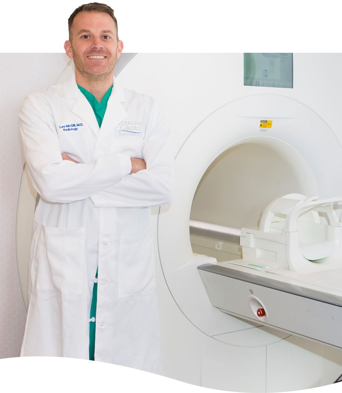
MRI is a non-invasive scan that uses a large magnet and radio waves to produce detailed images of almost every internal structure in the human body, including organs, bones, muscles and blood vessels. A 3T MRI scanner uses a stronger magnetic field than the standard 1.5T MRI scanner, allowing for clearer, more detailed images, faster scans and improved diagnoses, particularly for neurological and musculoskeletal conditions.
After-hours MRI services are now available Monday-Friday, 7 a.m. to 8 p.m. A physician’s order and insurance pre-certification are required. Call 229-985-EXAM (3926) to schedule or learn more.
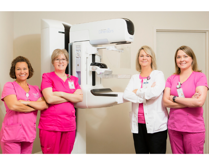
Mammography uses low-dose X-rays to examine your breasts, primarily for the early detection of breast cancer. Mammograms can be used for both screening and diagnostic purposes.
Screening mammograms can detect cancer before women experience symptoms, when it is most treatable. Schedule your mammogram today by requesting an appointment here or calling 229-985-3926.
Utilizing radioactive tracers with a small amount of radioactive substance injected into your bloodstream, nuclear medicine can visualize and assess organ function to diagnose and treat disease. A specialized camera tracks the path of the tracers.
Also known as a nuclear stress test, this procedure shows how blood flows to the heart at rest and during exercise. A small amount of radioactive material, called a tracer or radiotracer, is given by IV to enable images of the tracer moving through your coronary arteries. This helps to find areas of poor blood flow or damage.
This stress test can be used to help determine your risk for a heart attack or other cardiac event if your doctor knows or suspects that you have coronary artery disease. It may also be used to guide your treatment if you’ve been diagnosed with a heart condition.
This nuclear medicine imaging technique uses radioactive tracers to visualize your body’s metabolic processes. It helps your physician assess organ and tissue function, detect cancer and monitor the effectiveness of treatment.
Ultrasound is a safe and non-invasive imaging technique that uses high-frequency sound waves to create images of your organs and tissues. It is commonly used to diagnose and monitor a variety of medical conditions, including pregnancy, heart problems and blood clots.
These non-invasive procedures use sound waves (ultrasound) to assess blood flow in your arteries and veins. Essential for evaluating blood vessel health, detecting blockages and assessing treatment effectiveness, these studies help diagnose and monitor vascular conditions such as peripheral arterial disease, deep vein thrombosis and atherosclerosis.
Locations
Providers
Why a mammogram?
Early detection is key to a cure. A physician referral is not required for a screening mammogram at our state-of-the-art diagnostic imaging department, accredited by the American College of Radiology. 3D mammography and bone densitometry make access to care easy and convenient.
Request an appointment here or call 229-985-3926. Walk-ins are available for mammograms at Sterling Center Women’s Health

Request More Information
From X-rays to advanced MRI and CT scans, our expert imaging team delivers fast, accurate results with compassionate care. Contact us to get the clarity you need for your health.


